

3D MAMMO IS HERE!
What is 3D Mammography?
3D Mammography is an X-ray examination of the breasts. The value of mammography lies in its ability to detect cancer in the breast while it is still very small – often too small to be felt and often too small to be detected any other way. When breast tissue is x-rayed, it creates an image that may contain tiny calcium deposits called microcalcifications or other subtle signs of cancer. The Hologic 3D Tomosynthesis Mammography system provides a detailed image that can detect early breast cancer.
“I had a mammogram done by a friendly, caring professional. My primary doctor was impressed with the thorough report. I will be coming to River Radiology for my annual exam from now on!”
3D Mammography exam results are more accurate than 2D mammography alone. Conventional mammograms provide doctors with a two dimensional images of the three dimensional breast. This system provides the Radiologist with a series of detailed images allowing them to better evaluate your breast tissue layer by layer making fine details more visible and no longer hidden by overlapping tissue. This improved accuracy and can reduce false positive recalls for additional imaging.
When is Mammography Indicated for Screening?
If your doctor has suggested that you have a mammogram, it does not necessarily mean he or she suspects that you have breast cancer. The earlier cancer is found, the better the chances of a cure. That’s why the American Cancer Society and American College of Radiology recommend yearly screening mammograms starting at age 40 for women at average risk of breast cancer. Women at increased risk (family history, genetic tendency, past breast cancer, etc.) should talk with their doctors about the benefits and limitations of starting screening earlier, having additional tests, or having more frequent exams.
When is Mammography Indicated for Symptoms?
Women with new symptoms such as a lump, nipple discharge, or pain in the breast should see their doctor to determine if they should have a diagnostic mammogram. The mammogram can provide more information to help determine whether or not a biopsy is advisable. Sometimes your doctor will also recommend an ultrasound examination of the breast, which can complement the findings on a mammogram. It is important to remember that most lumps and other symptoms do not turn out to be cancer – even if a biopsy is recommended.
The 3D Mammography ProcedureThe process of a 3D exam is the same as a conventional 2D exam. The technologist will position you, compress your breast and take images from different angles. There is no additional compression required, and it only takes a few seconds longer for an exam proven to be more accurate. |
Your Mammography exam
Mammography is a quick and easy procedure. For a screening exam, please allow approximately 30 minutes. Allow an additional 30 minutes if an ultrasound or bone density exam is also scheduled. It is necessary to undress from the waist up for the exam and a gown will be provided. Your skin should be clean. Don’t use any deodorant, powder or lotion of any kind in your underarm or breast area, since the residue from such preparations can obscure your mammogram films. If you have deodorant on, we have wipes that you can use to take the deodorant off. Scripts are now required for all mammograms (diagnostic and screening). For more information you may visit http://www.cancer.gov/types/breast/mammograms-fact-sheet.
Remember - Mammography is not perfect!
While mammography is the single best method of detecting most breast cancers, it cannot find all cancers. Some cancers may be detectable only by physical examination. Some women may choose to do monthly breast self-examinations, as well as have annual exams by their doctor. If you or your doctor do feel something suspicious in your breast, remember that a normal mammogram cannot completely exclude the possibility of cancer – additional investigation (such as ultrasound, MRI, or biopsy) may be recommended. Remember also that most mammographic findings are not caused by cancer, even when additional testing or biopsy has been recommended.
When can my physician expect a copy of my report?
In most cases, reports are provided within two – three business days.
For women with a personal or strong family history of breast cancer, annual mammography may not be sufficient. That’s why River Radiology continues to invest in new technologies for breast cancer diagnosis.
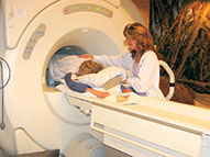
MRI Vibrant
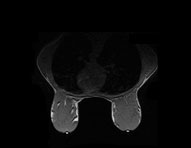
Computer-Aided Detection (CAD) for Breast MRI
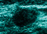
Ultrasound for Breast Imaging
For more information about breast cancer please visit www.acs.org

