
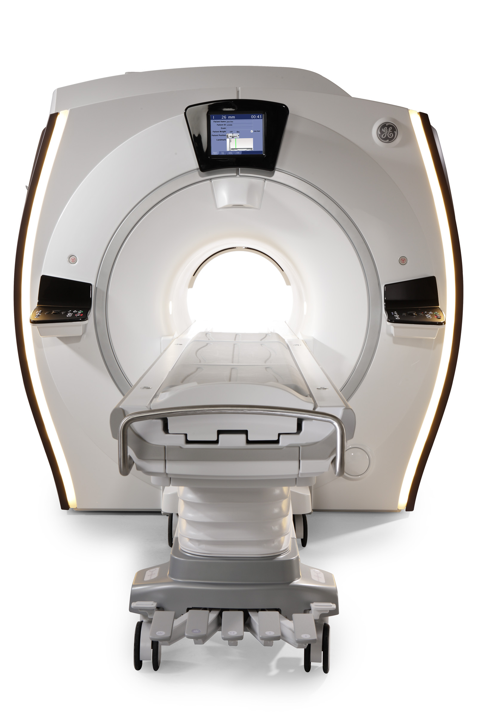
Our upgraded wide bore GE Optima MR 450w 1.5 T system with GEM Suite Technology is here!


What is MRI?
MRI is “magnetic resonance imaging.” MRI uses a powerful magnet, radio waves, and a computer to produce cross-sectional images and can be used to image almost any part of the body. No X-rays or other ionizing radiation are used.
Wide Bore MRI
The new wide bore MRI has a powerful magnetic field strength that is the one most preferred by physicians. It also makes scanning sessions faster. This wide MRI enables us to scan more anatomy in less time compared to previous generation systems, resulting in an improved scanning experience.
Wide bore refers to the shape of the opening that the patient lies in; the bore is not only wider but also shorter in length. Traditionally, wide bore scanners had limitations and poorer image quality. Our new wide bore MRI system improves image quality while allowing more room around the patient, providing more comfort during the examination.
“Some patients can present special challenges when undergoing MR testing,” said Dr.Susan Connors. “Now we can accommodate patients who weigh up to 500 pounds, and even those who don’t like small spaces can be more easily imaged and diagnosed with high quality scans.”
The newly installed and upgraded GE Optima 450 with GEM suite coil technology is designed to reduce scan times, reduce metal artifact around implants, decrease noise decibels, and correct image blurring due to motion, thus reducing the chance of recalls while delivering uncompromised imaging quality. The system also features a softer table to help maximize patient comfort and alleviate pressure points.
How does MRI work?
The physics and mathematics of MRI are extremely complicated. A large magnet surrounding the body causes the hydrogen nuclei (protons) in water molecules to align in a particular direction, and then radio waves strike the protons, causing them to spin out of alignment. When the radio signal is turned off, the protons return to their previous alignment, giving off new radio waves at the same time. The returning radio signals are received and analyzed by a computer, which then creates cross-sectional images of the body.
When is MRI used?
Like CT, MRI can be used to image almost any part of the body. MRI pictures contain information that may not be seen on a CT scan. For some things MRI is more useful than CT, and for some things CT is more useful. Sometimes, both are necessary, since the information gained from one may complement or clarify information gained from the other. MRI is often used for the following:
- Brain and nervous system: MRI is the most accurate tool for evaluating most diseases of the brain and spinal cord, including strokes, tumors, and multiple sclerosis.
- Musculoskeletal system: MRI is the most accurate way to non-invasively image most joints (knees, shoulders, wrists, etc) for problems involving cartilage, ligaments, tendons, and muscles.
- Breast: Both breasts are examined simultaneously in high resolution in a single patient visit.
- Other: MRI is useful for evaluating the neck, liver, kidneys, pelvic organs, blood vessels, and lymph nodes.
What is MRA?
MRA is “magnetic resonance angiography”. Using special techniques, MRI can be employed to produce highly detailed images of many arteries and veins, without the need to insert catheters into the body or inject iodinated contrast agents. The pictures obtained look much like a conventional angiogram, but are obtained non-invasively.
Preparing for an MRI at our facility
Not everyone can be safely exposed to a magnetic field, so if any of the following apply to you, you should let us know before making an appointment:
- cardiac pacemaker
- history or possibility of a metal fragment in your eye, even if it has been removed
- aneurysm clip in the brain
- ear or eye implant
- nerve stimulator
- drug infusapump
- recent surgery (less than 8 weeks ago)
- any other procedure where medical personnel might have left a stent, coil, filter, wire, or any other implantable device inside your body
- Please also let us know if you weigh more than 450 lbs, or if there is any possibility that you may be pregnant.
Allow 1 to 1-1⁄2 hours for the appointment. Patients must wear metal free clothing such as jogging or sweat pants with elastic waists and no zippers. Do not wear any metal infused sportswear. There may be eating or drinking restrictions for abdomen and/or pelvis exams. Because MRI uses a powerful magnet, patients can not have any metal on or in their bodies, including piercing, aneurysm clips, or stents. If there is any possibility that you have metal in or around your eyes, it is necessary that we schedule an X-ray of your eyes, prior to the MRI. A nurse or technologist will conduct a thorough interview prior to the exam. As with all imaging procedures please bring prior images, a script, and your insurance card(s) with you. (NOTE: Patients with pacemakers cannot have an MRI at our facility.)
You will be positioned on a comfortable scanning table. Some examinations require the injection of a small amount of Gadolinium-contining contrast — a substance that improves the visualization of some structures and is necessary to demonstrate some diseases or conditions (especially for some brain and abdominal exams). This substance almost never causes side effects, and is quickly excreted by the kidneys. A “surface coil” may be placed on or around the part of your body to be imaged, such as the head, knee or shoulder. Since the large magnet and rapidly changing magnetic field produce loud noises at times during the exam, you will also be given ear protection. Most exams take anywhere from 15 to 45 minutes to complete. Prior to the exam room, a staff member will interview you and explain what will happen during your test. A technologist will stay in constant contact with you throughout the exam
When can my physician expect a copy of my report?
Most reports are available within 1 business day or less, and are accessible through our Patient Portal.


