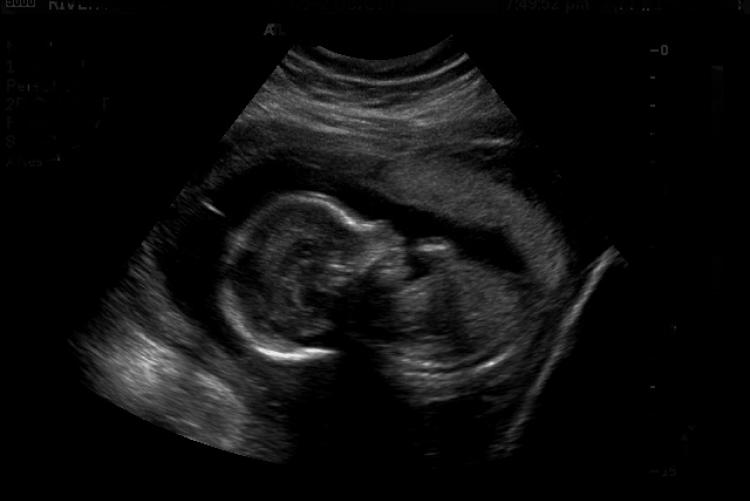
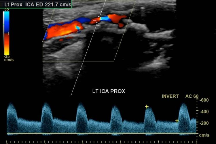
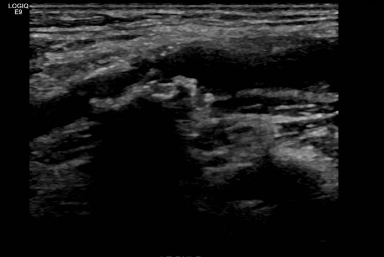
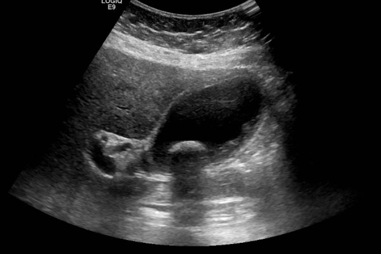
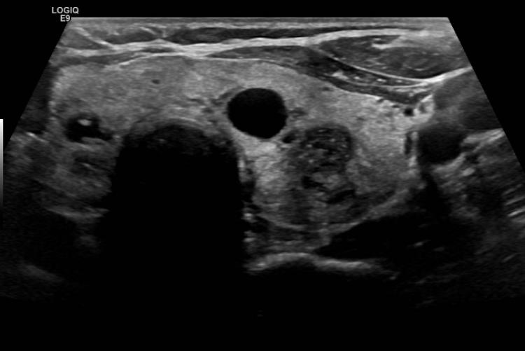
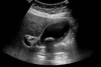
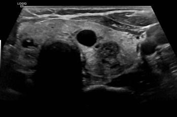
Ultrasound imaging, or sonography, is a painless non-invasive and expedient way to look inside the body at organs and soft tissue. There is no exposure to ionizing radiation as there is with X-ray or Computed Tomography (CT) exams. Instead, ultrasound uses high frequency sound waves to create a detailed image of the organ or tissue being examined. Sound waves are sent into the body through a hand-held device called a transducer which is pressed against and moved over the skin. The ‘echoes’ that bounce back are then displayed as an image in real time on the ultrasound monitor.
River Radiology has recently acquired the GE XDclear ultrasound system. This premium system provides innovative technologies to help improve patient care. The XDclear™ transducer is advanced ultrasound transducer technology from GE Healthcare that is enhancing the scanning of obese or bariatric patients. The XDclear transducer generates a high quality ultrasound signal to provide deep sound wave penetration in order to achieve high definition resolution throughout the image.
What is an Ultrasound?
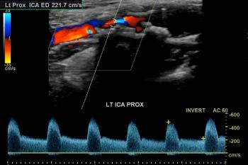
Diagnostic Ultrasound is the use of high-frequency sound waves to obtain images of the body. The sound waves can be used to produce images of various organs, to observe motion within organs, or to measure the flow of blood using a specialized method called Doppler Ultrasound.
How does Ultrasound work?
A small, hand-held device, called a transducer, is used to send sound waves into the body, and to receive returning signals. The information returning to the transducer is sent to a computer, which reconstructs cross-sectional images of the body – similar in concept to sonar or radar.
When is Ultrasound used?
Because Ultrasound is non-invasive, painless, and does not use ionizing radiation, it is an extremely valuable way to evaluate many parts of the body – including the pregnant uterus, pelvic organs, kidneys, gallbladder, liver, thyroid, breasts and testicles. Doppler Ultrasound is used to evaluate the heart, as well as blood vessels such as the carotid arteries and veins of the legs.
Preparing for an Ultrasound exam
There are different preparations for different kinds of exams.
- Pregnancy, pelvic, and bladder sonogram:
You must have a full bladder for these exams
**We understand the desire to be in the room with the patient for a pregnancy ultrasound but to ensure that the sonographer can focus completely, we will ask that only the patient be in the room**
- Abdomen, gallbladder, liver and kidney sonograms:
No solid foods for 6 hours prior to your exam.
The exam is performed by a specially trained Ultrasonographer. Most ultrasound exams (of the kidney, bladder, pelvis, fetus, abdomen and liver) require patient preparation, including drinking water prior to the exam. Allow 30 to 60 minutes for the appointment depending on the specific exam. The exam room is usually darkened to improve the visibility of the video monitor.
The Ultrasound examination
The procedure is painless. You will lie on a comfortable exam table, while the sonographer moves a transducer over your skin. A gel-like substance is applied to the skin, to allow the sound waves to pass through more easily.
Why do some exams require me to have a full bladder?
A full bladder acts as a “window” in an otherwise solid “wall,” the abdomen/pelvis. This enables us to look through the window to what lies behind it, including the uterus and ovaries.
When can my physician expect a copy of my report?
Most reports are available within a week and are accessible through our Patient Portal 4 business days after it has been read.

